
Introduction
The mechanisms underlying human erection constitute a complex and intricate network in which central, hormonal, neurological, arterial, and venous factors interact. The relative weight of each component is variable; however, penile veno-occlusive dysfunction (VOD) is increasingly recognized as an important cause of erectile dysfunction in patients under 40 years of age.1
Recent studies have suggested an association between penile ultrastructure—particularly the architecture of the tunica albuginea (TA) or the venous wall—and the occurrence of venous leakage. This makes VOD an emerging etiological field under systematic investigation, with novel therapeutic approaches being explored and several research questions still to be answered.2
The cornerstone of successful surgical management lies in the careful selection of patients. The renewed interest in venous leak disorders has been largely driven by the advent of more refined diagnostic methods, such as high-definition penile ultrasound and computed tomography-cavernosography (Caverno-CT), which allow for better correlation with clinical findings and more accurate identification of patients who may benefit from surgical treatment.3,4
History of venous leak surgery
More than 100 years of history have not sufficed to achieve a comprehensive understanding of venous leakage. Surgery for venous leaks dates back at least to the 19th century, with Raymond and Duncan in 1895.5 Since then, enthusiasm for these procedures has fluctuated significantly. Venous ligation surgery was reported as early as the beginning of the 20th century by Wooten,5 who described it as a cure for so-called atonic penis. The technique was later revisited by various authors until its peak in the 1980s, when numerous studies were published, including those by Wespes and Shulman6,7, Lazzarini et al.8, Shabsigh et al.9 and Lue et al.,10. The central problem was highlighted by Trost et al.11, who noted the scarcity of relevant literature on penile venous surgery and concluded that, with the available data, it is not possible to establish standardized criteria for patient selection, hemodynamic testing, surgical technique, follow-up, or definitive outcomes. A renaissance of the pathophysiological concept and the proposal of new surgical techniques emerged with the contributions of Korean urologist Hsu et al.12–14, which are discussed in this article. Likewise, the team at Hospital Pablo Tobón Uribe in Medellín has conducted continuous research and published several studies on a laparoscopic correction technique, which is also extensively reviewed herein.3,15,16
Pathophysiology of penile venous leaks
Two types of dorsal venous leakage may occur: primary (congenital) and secondary (acquired). Secondary leaks, which are associated with various penile pathologies that produce acquired venous leakage at different sites, are not covered in this review. Regarding congenital dorsal venous leaks, their pathophysiology has been debated for more than a century, with no single theory universally accepted17. One hypothesis suggests an intrinsic defect in the venous wall, like that observed in patients with varicocele or other peripheral venous disorders (e.g., varicose veins, hemorrhoids), leading to insufficiency from the onset of sexual life (primary erectile dysfunction [PED]) or before the age of 40 (non-standard erectile dysfunction). Another hypothesis attributes venous leakage to alterations of surrounding tissue, particularly the TA. In this model, the subtunical venular plexus and emissary veins traverse the tunic between sinusoidal spaces, allowing compression and occlusion during erection. As smooth muscle relaxes and the sinusoids expand with increased blood inflow against the fibrous-elastic tunica, blood becomes trapped within the corpora cavernosa.2,3,18,19
Patient profile: deep dorsal venous leak syndrome
Although Campbell’s Urology20, the AUA guidelines for erectile dysfunction, and the standard international society for sexual medicine practice in sexual medicine21 do not recommend surgical correction of penile venous leaks, recent publications suggest that penile venous surgery for congenital deep dorsal venous leakage may yield excellent long-term results and patient satisfaction in a highly selected population of young men. PED or non-standard erectile dysfunction (onset before 40 years of age)4 represents a diagnostic challenge, as these patients may be candidates for curative surgical intervention. Young men with erectile dysfunction, particularly those presenting with easy loss of erection or a wavering erection pattern (WEP) – that is, erectile failure in situations of reduced arousal or concentration – should be evaluated for possible congenital venous leakage.
The so-called Deep Dorsal Venous Leak Syndrome includes the following components:
- – PED or non-standard erectile dysfunction of more than 6 months’ duration before the age of 40
- – Presence of a WEP: Soft erections of short duration, easily lost with minimal reduction in sexual stimulus, under conditions such as:
- Condom application.
- Change of position.
- External distractions (noise, interruptions, phone use, etc.).
- Erection loss while standing (most common), although ~20% of patients exhibit the inverse pattern (greater loss while supine).
- Persistence of WEP during masturbation.22
- – ≥ 75% erection achieved with vasoactive stimulation during pharmacological duplex Doppler ultrasound, ruling out inadequate stimulation as a confounding factor
- – Diagnosis of venous leak: confirmed deep dorsal venous leakage on penile ultrasound and/or Caverno-CT, with no evidence of additional venous leaks in the corpora cavernosa (Fig. 1).
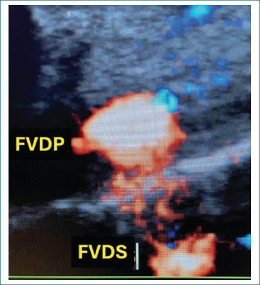
Figure 1. Deep dorsal venous leakage and superficial dorsal venous leakage in Doppler ultrasound HD (courtesy of the authors).
Diagnosis of venous leaks
Penile Doppler ultrasound
Penile Doppler ultrasound is a cost-effective tool for the evaluation of penile hemodynamics, providing information on both the arterial system and the presence of venous leaks, which can be localized during the different phases of erection. The diagnostic criteria for veno-occlusive disease on penile Doppler ultrasound are:
- – Peak systolic velocity > 30 cm/s (normal arterial function)End-diastolic velocity of 0 or very close to 0
- – Continuous record of blood outflow (leakage) through the deep dorsal vein of the penis throughout the entire examination, with sufficient penile tumescence
- – No other cavernous venous leaks. Periurethral (spongiosal) leaks may be present but do not affect erectile hemodynamics.23–26
Digital cavernosography
An alternative diagnostic method involves the intracavernous injection of contrast medium with sequential radiographic imaging to detect venous leakage. Pharmacologically induced erection or, at a minimum, erection with saline solution is a prerequisite. Currently, this study is performed using digital radiology systems, which allow video sequences of the injection (Fig. 2).27,28
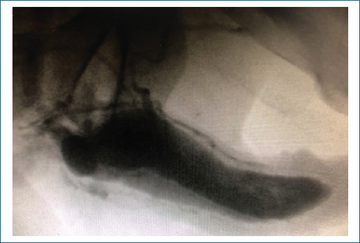
Figure 2. Deep dorsal venous leak in oblique view during cavernosography with iodinated contrast medium (courtesy of the authors).
Caverno-CT
The use of computed tomography with artificial erection and intracavernous contrast injection can markedly improve image quality. This technique provides three-dimensional visualization, rotation, color enhancement, image reconstruction, and tissue subtraction (e.g., bone), all of which increase diagnostic accuracy. Proper execution requires collaboration between a urologist and a radiologist trained in the procedure (Fig. 3).26,29,30
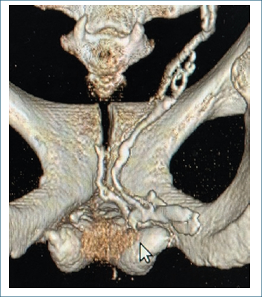
Figure 3. Deep dorsal venous leakage to the Santorini plexus on computed tomography cavernosography.
Treatment options for dorsal venous leaks
Non-surgical options
ORAL PDE5 INHIBITORS
Vasodilator therapy with PDE5 inhibitors enhances arterial inflow, leading to transient improvement of erectile function without addressing the underlying pathology. Sildenafil, tadalafil, avanafil, and vardenafil have all been used, either on demand, daily (tadalafil), or in combination (short-acting on demand plus daily dosing). In our series of 20 patients with dorsal venous leak and moderate-to-severe ED, only 2 improved to mild ED after 1 year of medical therapy, with no cases of full recovery.28
INTRACAVERNOUS VASOACTIVE THERAPY
Similar to PDE5 inhibitors, vasoactive injections alter the inflow-outflow balance. Alprostadil or combination regimens can improve erection quality, but escalating doses are often required, raising the risk of prolonged erections. This is a palliative option.3
FLOW-CONTROLLING CONSTRICTION RINGS
Mechanical occlusion at the penile base may help limit venous outflow. This option requires strong patient and partner adherence.3
VACUUM ERECTION DEVICES (VEDS)
VEDs can complement constriction rings and, in adherent patients, serve as effective inducers of passive erections.3
LOW-INTENSITY SHOCKWAVE THERAPY (LI-ESWT)
Although promising in vasculogenic ED of arterial origin, LI-ESWT is ineffective in venogenic ED, where the underlying mechanism is venous leakage. Nevertheless, it has been proposed as a potential adjunct therapy.31
Surgical options
Open venous ligation
The classic surgical option involves selective exposure of the dorsal veins through circumcision or dorsal penile incision. The most widely used current technique is venous stripping, proposed by Korean andrologist Hsu et al. This technically demanding surgery requires isolation and ligation of all major veins – including circumflex, oblique, perforating, and even the smallest branches – along the penile shaft.12,32–39
Laparoscopic venous ligation
A variation involves laparoscopic access to the Santorini plexus, where the penile dorsal veins converge. The technique resembles an initial hemostatic step in radical prostatectomy. The procedure is described in detail in this article.27,28
Dorsal vein embolization
Interventional radiology has proposed selective embolization of the dorsal veins using agents such as polidocanol (aethoxysklerol), N-butyl cyanoacrylate, endovascular coils, or combinations thereof.40–42
Device-assisted venous ligation
Under investigation are devices designed for the implantation of nitinol coils through an open, minimally invasive penile approach.
Penile prosthesis implantation
As in other forms of ED, penile prosthesis implantation remains a last-resort treatment. Given that most patients with venous leak are very young, implants should not be proposed without thorough evaluation and consideration of other alternatives, such as laparoscopic correction.
Step-by-step laparoscopic venous ligation technique
- – Step 1. Patient positioning: supine with dorsal elevation using a roll or cushion (Fig. 4).
- – Step 2. Initial incision: a midline subumbilical incision is made after placing a Foley catheter (No. 14) to empty the bladder (Fig. 5).
- – Step 3. Dissection into Retzius space (with or without balloon): Preferably extraperitoneal. Dissection begins subcutaneously with digital access, using one of the following:
- – Step 4. Pneumoretroperitoneum parameters: CO2 pressure set at 15-20 mmHg; electrocautery in pure coagulation mode at 40 W (Fig. 8).
- – Step 5. Trocar placement: central 10-mm trocar for the camera, directed obliquely to Retzius space, followed by CO2 insufflation and insertion of two lateral 5-mm trocars under direct vision. Incidental pneumoscrotum may occur but has no clinical consequences (Fig. 9).
- – Step 6. Periprostatic dissection: areolar and fatty tissue in the periprostatic region and recesses are dissected, exposing the Santorini plexus, usually with a hook cautery (Fig. 10).
- – Step 7. Santorini plexus dissection: adequate visualization of the Santorini plexus is the goal (Figs. 11 and 12).
- – Step 8. Santorini plexus ligation: achieved with a non-absorbable Ethibond 0 suture passed through the 10-mm trocar and tied with a double knot. It should be noted that ligation is performed exclusively on the venous bundle of the Santorini plexus. (Figs. 13 and 14).
- – Step 9. Accessory vein ligation: If present, accessory veins are clipped with titanium ligaclips or disposable polypropylene Hem-o-Lok clips. Passive penile congestion induced with a digital tourniquet is recommended to localize accessory veins (Fig. 15).
- – Step 10. Final steps: Skin closure with Prolene (non-absorbable) or Monocryl (absorbable). Local bupivacaine blocks at incision sites. Removal of Foley catheter, evacuation of pneumoscrotum if present, and same-day discharge with 3-5 days of recovery.
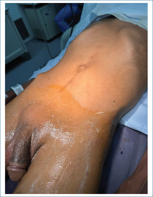
Figure 4. Patient position supine with dorsal lift.
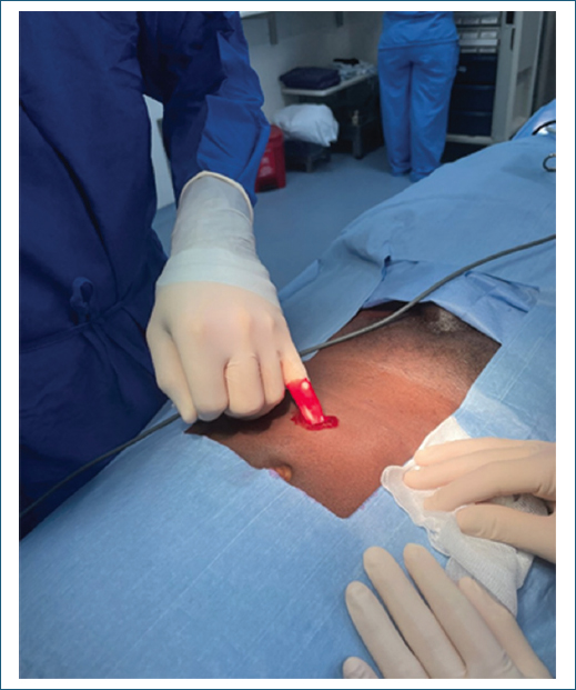
Figure 5. Initial incision for dissection of the Retzius space for a 10 mm trocar.
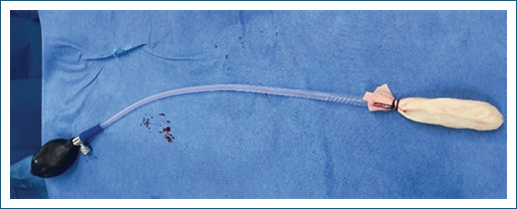
Figure 6. Device created for the dissection of the Retzius space and its use.
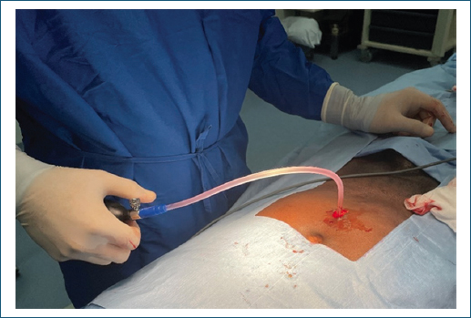
Figure 7. Device created for the dissection of the Retzius space and its use.
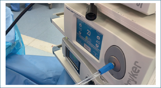
Figure 8. CO2 equipment parameters.
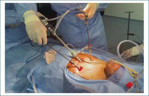
Figure 9. Position of the three trocars.
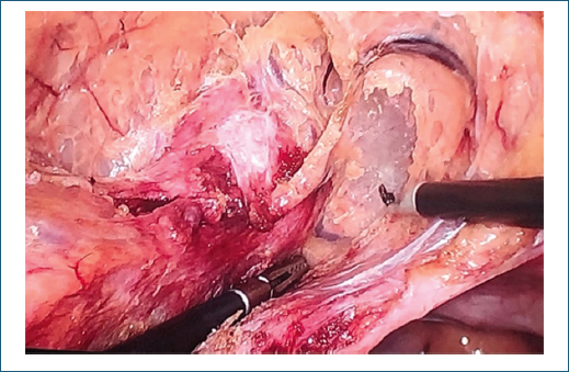
Figure 10. Periprostatic dissection and recesses.
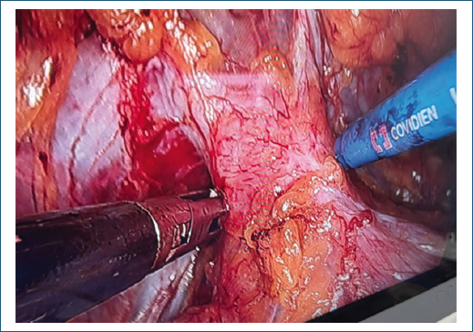
Figure 11. Dissection of Santorini’s plexus.
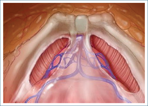
Figure 12. Illustration of Santorini plexus (modified from: https://link.springer.com/chapter/10.1007/978-3-319-32641-2_3).
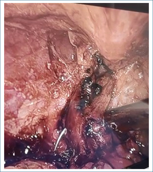
Figure 13. Ligation of Santorini’s plexus.
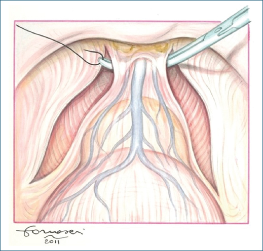
Figure 14. Illustration of ligation of Santorini Plexus (modified from: Cristini et al. BMC Urology 2013, 13:13 http://www.biomedcentral.com/1471-2490/13/13).
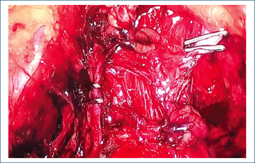
Figure 15. Ligation of accessory veins with clips.
Complications
- – Complications related to laparoscopic access: bleeding, vascular injury, and adjacent organ injury (e.g., bladder).
- – Late complications related to laparoscopy: wound dehiscence and incisional hernia.
- – Complications related to ligation of the Santorini plexus: adjacent organ injury (e.g., urethral injury).
- – Procedure-related complications: pain and infection
- – Catheter-related complications: dysuria and urinary retention.
Conclusions
When a deep dorsal venous leak is suspected in patients under 40 years of age, associated with hemodynamic alterations explaining a specific type of erectile dysfunction, the entity known as deep dorsal vein leak syndrome should be considered. This condition is characterized by clinical findings dominated by the WEP and ultrasonographic confirmation of deep dorsal venous leakage in the setting of preserved arterial function, corroborated by complementary tools such as Caverno-CT.
Although medical alternatives exist, laparoscopic surgical correction of venous leakage is proposed as a viable and reproducible therapeutic alternative. The technique is simple, reproducible, accessible to any laparoscopically trained team, associated with minimal complications, favorable erectile outcomes, preservation of penile anatomy, outpatient feasibility, and short recovery time.
Recommendations
Further studies and collaboration among multiple professional groups performing and reporting on this technique are required to consolidate the evidence and optimize the management of this pathology.
These findings are limited by the fact that they reflect the surgical experience of a single group of authors at one institution; therefore, it is mandatory that other groups publish their experience with the same technique.
Funding
The authors declare that this work was carried out with the authors’ own resources.
Conflicts of interest
The authors declare that they have no conflicts of interest.
Ethical considerations
Protection of human subjects and animals. The authors declare that no experiments on humans or animals were performed for this research.
Confidentiality, informed consent, and ethical approval. The authors have followed their institution’s confidentiality protocols, obtained informed consent from all patients, and secured approval from the Ethics Committee. SAGER guidelines have been followed as applicable to the nature of the study.
Declaration on the use of artificial intelligence. The authors declare that no generative artificial intelligence was used in the writing or creation of the content of this manuscript.
